Kolegij
Studiji
Medical Studies in EnglishStudijska godina
2ISVU ID
187245ECTS
5
ECTS grading system: Student grading will be conducted according to the current Ordinance on Studies of the University of Rijeka and the Ordinance on Student Grading at the Faculty of Medicine in Rijeka. Student work and achievement are assessed and graded during the course, which is the basis for the final grade. Student work and competencies are evaluated during classes with a maximum of 70 grade points and up to 30 grade points at the final exam, which totals 100 grade points. Students are graded according to the ECTS (A-E) and numerical system (1-5). Grading according to the ECTS system is conducted according to the absolute redistribution, as well as according to the graduate grading criteria.
I. The following components are evaluated during the course (maximum of 70 grade points):
a) acquired knowledge (up to 66 grade points)
b) seminar thesis (up to 6 grade points)
a) acquired knowledge (up to 66 grade points)
During classes, acquired knowledge will be evaluated by two midterm exams comprising 60 questions, which will take place on November 2020 (first midterm exam) and on January, 2021 (second midterm exam). A student may obtain up to 16,5 grade points on each midterm exam:
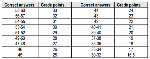
Students who fail to earn a minimum number of points one or both MTEs can repeat one or both MTEs, which will be organised in February, between the first and second term of the Final exam. At repeated MTEs, student can acquire grade point according to the above table and correct/improve the final score.
Improvement of the overall performance during the course. Students who have achieved sufficient points on a regular MTEs can improve their final score at the repeated MTE/MTEs. The repeated MTEs (writing the test) will be organized at the Faculty of Medicine under controlled conditions: either using traditional printed tests or using the Merlin platform in the faculty's computer classroom.
Additional acquisition of minimum conditions for the Final exam. Students who failed to acguire a minimum score on one of the MTEs can earn minimum grades required to access the Final exam. This will be organized in early September. The acquisition of minimum grade pointe will be carried out by writing one or both tests covering the material of the first and/or second MTE. The acquisition of minimum garde points (writing a test) will be organized at the Faculty of Medicine under controlled conditions: either using traditional printed tests or using the Merlin platform in the Faculty's computer classroom. On tests for the acquisition of minimum conditions, students cannot earn additional grade points With a positive test result (more than 50%) student can earn the minimum number of grade points (17.5+17.5) and can access the Final exam. If it is not possible to approach the Faculty due to the epidemiologic situation, additional acquisition of minimum conditions will be carried out by oral examination of the required materials using MS teams or Google Meets. At the oral check, students can achieve a positive result and earn the minimum number of points needed to enter the Final exam.
b) Independent work (up to 4 grade points)
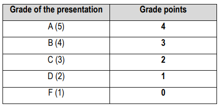
A student must prepare a Powerpoint presentation and present it to other students during practicals (starting from P2). After that, a student submits the presentation to the teacher in a printed form with a front page containing the topic title, name and surname of the student, their group, and the date. The presentation should not last longer than 10 minutes, and the student can choose only one topic for the presentation. The list of topics will be announced at the Share-portal of the course. The number of grade points granted for the presentation is evaluated by the teacher according to the quality of content and presentation in categories.
A positively evaluated presentation in a certain field is graded as follows:
Attending lectures, seminars, and practicals are mandatory. Students can be absent from 30% of classes provided they have a justifiable cause, i.e. a doctor’s note. If a student is absent for more than 30% of the classes, whether it is justifiable or not, they cannot continue to participate in the course and cannot access the final exam. In that case, the student is graded with 0 ECTS points and an F grade.
II. Final exam (up to 30 grade points)
Students who obtained 35-70 grade points during classes are obligated to access the final exam at which they may obtain additional grade points. The final exam consists of a multiple-choice questions test and an oral part. Students who obtained less than 35 grade points during classes or were absent for more than 30% of classes are not allowed to access the final exam (insufficient F).
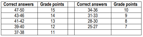
Students can obtain 15-30 grade points at the final exam. The final exam consists of an oral and a written part, where students are expected to show at least 50% of knowledge, skills, and competencies. A student who demonstrates at least 50% of knowledge, skills, and competencies at the written and the oral part of the exam, is credited with points according to the achieved result, which is added to the grade points obtained during classes. At the written part of the final exam, a student can obtain 15 grade points according to the table:
At the oral part of the final exam, a student can obtain 15 grade points that are divided into 5 categories:
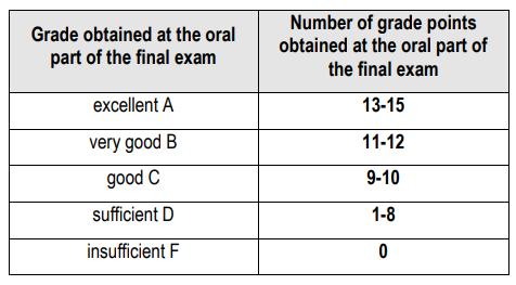
In order to pass the final exam, a student must achieve a minimum of 7 grade points at the written part and a minimum of 8 grade points at the oral part of the exam. The final exam is an integral part, therefore, if the student does not achieve a positive assessment of the oral part of the final exam, the results of the written part of the final exam are invalid in the following final exam terms.
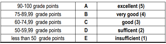
III. The final grade (maximum of 100 grade points) The final grade represents a sum of all grade points obtained during classes and at the final exam based on the absolute redistribution according to the following scale
Ishodi učenja
After successfully mastering the material, the student will be able to:
- Define the concept of homeostasis and the importance of maintaining the stability of the internal environment (extracellular fluid).
- Explain the differences between negative feedback, positive feedback, and feed-forward control in the regulation of physiological processes.
- Describe the concept of diurnal (circadian) rhythms and their influence on the variability of physiological values.
- Recognize the concept of hysteresis as a phenomenon where the state of a system depends on its history.
- Demonstrate examples of the physiological interplay of different feedback loops in the systemic integration of the organism.
- Apply the concept of a "tipping point" to predict when physiological compensation ceases to be sufficient.
- Illustrate the difference between an acute response to a stimulus and chronic adaptation, which shifts the boundaries of homeostasis.
- Analyze disease as a pathophysiological disruption of homeostatic mechanisms.
- Compare the efficiency and biological purpose of negative versus positive feedback in maintaining the stability of the organism.
- Critically evaluate the limits of physiological compensation under conditions of chronic stress or pathological changes in the internal environment.
Ishodi učenja
After this lecture, the student will be able to:
- Define the concepts of passive transport (diffusion) and active transport across the cell membrane.
- List the subtypes of passive transport (simple diffusion through the bilayer, pores/aquaporins, and channels; facilitated diffusion) and active transport (primary and secondary active transport).
- Identify the key transport proteins (aquaporins, GLUT transporters, ion pumps, symport, and antiport carriers).
- Explain the role of the chemical (concentration) gradient as the driving force for passive transport, contrasted with the energy (ATP) requirement of active transport.
- Describe the mechanism of facilitated diffusion using the GLUT transporter model (conformational change of the carrier without energy expenditure).
- Clarify the physiological distinction between primary active transport and secondary active transport.
- Describe the difference between symport (co-transport) and antiport (counter-transport/exchangers) using concrete physiological examples.
- Predict the direction and type of transport for a given substance (e.g., oxygen, glucose, water, ions) based on its physicochemical properties (lipophilicity, charge, size) and concentration distribution across the membrane.
- Apply the concept of carrier saturation (saturation kinetics /
 ) to distinguish the linear dynamics of simple diffusion from the saturating dynamics of facilitated diffusion.
 - Compare and contrast simple diffusion, facilitated diffusion, and active transport based on the following criteria: direction relative to the gradient, requirement of a specific protein, energy consumption, and susceptibility to saturation.
- Analyze how the pharmacological or pathophysiological inhibition of primary active transport (e.g., blocking the Na+/K+ pump) consequently impairs the function of secondary active transporters within the cell.
Ishodi učenja
After this the student will be able to:
- List the main types of ion channels based on selectivity (Na+, K+, Ca2+, Cl-) and gating mechanisms (voltage-gated channels and leak / semi-leaking channels).
- State the typical values of the resting membrane potential for a general model of a human cell.
- Explain the physical meaning of the Nernst potential as a state where the chemical (concentration) gradient and the electrical gradient for a single ion are in a dynamic equilibrium.
- Describe how selective membrane permeability (primarily through potassium leak channels) and the action of the electrogenic Ca2+ pump work together to generate the resting membrane potential.
- Apply the concept of the Nernst potential to predict the direction of ion movement (influx or efflux) if the actual membrane potential deviates from the equilibrium potential.
- Compare and contrast the role of continuously open leak channels, which dominate at rest, with the role of voltage-gated channels, which are mostly closed at rest.
- Correlate the high relative membrane permeability to potassium at rest with the fact that the overall resting membrane potential is very close to the Nernst potential for potassium (Ca2+).
- Critically assess the importance of continuous metabolic energy expenditure (ATP) for maintaining the ionic gradients, without which the resting potential would collapse.
Ishodi učenja
After this lecture, the student will be able to:
- Define the action potential (AP), threshold potential, the "all-or-none" law, and membrane refractoriness.
- List and chronologically arrange the phases of the action potential (depolarization, repolarization, hyperpolarization).
- Identify the states of hypo- and hyperkalemia, hypo- and hypercalcemia, and hypo- and hypernatremia based on ion concentration alterations in the extracellular fluid.
- Define the term channelopathies as primary disorders of ion channels.
- Explain the ionic mechanisms underlying the phases of the AP (the gating kinetics of activation, closure, and inactivation of voltage-gated Na+ and K+ channels).
- Clarify why hyperkalemia causes initial membrane depolarization but chronically leads to the inactivation of Na+ channels and a loss of excitability.
- Explain the role of extracellular calcium as a "membrane stabilizer" through its effect on the activation threshold of voltage-gated Na+ channels.
- Describe how alterations in sodium concentration affect the osmotic gradient, cell volume, and the amplitude of the action potential upstroke.
- Predict alterations in the shape, amplitude, and slope of the action potential waveform under conditions of severe hyponatremia or hypernatremia.
- Apply the concepts of absolute and relative refractory periods to explain the unidirectional propagation of the action potential along the membrane and the maximum firing frequency of impulses.
- Analyze and contrast the effects of hypokalemia and hyperkalemia on the resting membrane potential and its distance from the threshold potential (excitability).
- Correlate the pathophysiological mechanism of hypocalcemia (lowering the Na+ channel activation threshold closer to the resting potential) with the clinical manifestation of spontaneous muscle contractions (tetany).
- Differentiate secondary electrophysiological disorders (caused by electrolyte imbalances) from primary genetic defects of ion channels (channelopathies).
Ishodi učenja
After successfully mastering the material, the student will be able to:
- Explain the main cellular homeostatic mechanisms, including the regulation of pH, ionic balance (Ca²⁺), and volume maintenance.
- Define the concept of proteostasis and the role of chaperones in ensuring proper protein folding.
- Describe the function of key cellular "sensors" and "controllers" (DDR, UPR) in recognizing protein and DNA damage.
- Recognize the basic metabolic pathways through which the cell maintains energy balance under normal and hypoxic conditions.
- Demonstrate how the cell detects and responds to stress (e.g., the accumulation of misfolded proteins via the UPR pathway).
- Apply knowledge of second messengers and signaling pathways to explain the integration of external signals into the intracellular response.
- Illustrate the phenomenon of metabolic reprogramming (e.g., the switch to anaerobic metabolism) under conditions of limited oxygen availability.
- Explain the importance of contact inhibition and adhesion for tissue organization and the prevention of pathological (tumor) growth.
- Analyze the causes and consequences of cellular homeostasis disruptions, linking them to the development of cellular stress.
- Compare the roles of DDR (DNA Damage Response) and UPR (Unfolded Protein Response) in the cell's decision between survival and programmed cell death.
- Synthesize knowledge of energy homeostasis to assess how systemic hypoxia affects the function of individual organs and tissues.
- Critically evaluate how the collapse of proteostasis contributes to the development of degenerative diseases.
Ishodi učenja
After successfully mastering the material, the student will be able to:
- Explain the fundamental processes of maintaining cellular homeostasis, including the regulation of pH, calcium concentration, and energy balance.
- Define the concept of proteostasis and explain the role of chaperones in ensuring the integrity of the cellular proteome.
- Recognize various forms of cellular stress (oxidative, ER stress, genotoxic, and metabolic stress) and their characteristic biomarkers.
- Describe the role of sensors and control mechanisms (e.g., DDR, UPR) in recognizing and responding to cellular stress.
- Demonstrate how "metabolic reprogramming" enables cellular survival under conditions of limited resources or hypoxia.
- Apply knowledge of signaling pathways to explain how cells integrate external stimuli with intracellular responses.
- Illustrate the consequences of impaired contact inhibition and adhesion on cellular organization.
- Analyze the link between prolonged (chronic) cellular stress and the loss of cellular function or the development of pathological states.
- Compare acute stress response mechanisms (e.g., temporary adaptation) with permanent changes in cellular phenotype.
- Synthesize information regarding cellular sensor systems to explain how a cell "decides" between repair processes (e.g., DDR response) and programmed cell death.
- Critically evaluate the importance of redox homeostasis and the impact of its disruption on overall cellular pathophysiology.
Ishodi učenja
After successfully mastering the material, the student will be able to:
- Define the basic forms of cellular adaptation to stress (atrophy, hypertrophy, hyperplasia, metaplasia).
- Explain the fundamental differences between apoptosis (programmed death) and necrosis (acute, uncontrolled death) and their respective molecular triggers.
- Describe the phenomenon of autophagy as a process that balances between maintaining cellular homeostasis (survival) and directing the cell toward death.
- Recognize the role of stem cells as the fundamental reservoir for the regenerative capacity of tissues.
- Demonstrate the cascade of events during ischemia-reperfusion injury and explain why reperfusion can further damage tissue.
- Apply the understanding of necroptosis as "programmed necrosis" in explaining certain inflammatory processes.
- Illustrate how the free radical theory links oxidative stress to the process of cellular aging (senescence).
- Analyze the signaling cascades that determine whether a stressed cell will survive, adapt, or be removed by apoptosis.
- Compare different mechanisms of cell death (apoptosis, necrosis, necroptosis) in the context of clinical consequences for the organism (e.g., inflammatory response vs. silent cell elimination).
- Synthesize knowledge regarding the function of stem cells to evaluate the potentials and limitations of regenerative medicine in treating chronic degenerative diseases.
- Critically evaluate the biological role of cellular aging (senescence) as a protective mechanism against the tumor transformation of cells.
Ishodi učenja
After successfully mastering the material, the student will be able to:
- Define the fundamental characteristics of tumor cells (the "hallmarks of cancer") that enable uncontrolled proliferation and survival.
- Explain the role of genomic instability in initiating tumor transformation.
- Describe the processes of angiogenesis (formation of new blood vessels) and metastasis as key factors in tumor progression.
- Recognize the importance of immune surveillance and the mechanisms by which tumor cells successfully evade it.
- Demonstrate tumor growth kinetics and explain the difference between the rate of cell division and the total volumetric increase of the tumor.
- Apply knowledge of cellular metabolism to explain the concept of "overcoming the energy barrier" in tumor cells.
- Illustrate the clinical significance of tumor cell differentiation in establishing a diagnosis and assessing disease prognosis.
- Analyze biological error as a process that leads from mutation to a clinical entity (tumor).
- Compare invasive growth with normal tissue adhesion and cell movement.
- Critically evaluate the translational aspects of tumor biology, with a particular focus on the possibilities of gene therapy and personalized medicine.
- Synthesize theoretical knowledge of tumor molecular biology by participating in a simulated "Molecular Tumor Board," linking the tumor's genetic profile to therapeutic strategies.
Ishodi učenja
After successfully mastering the material, the student will be able to:
- Define the role of muscle as an electromechanical transducer in the context of converting ionic currents into mechanical force.
- Describe the architecture of the sarcomere, identifying key protein components (actin, myosin, troponin, tropomyosin) as the molecular blueprint of contraction.
- Explain the role of the triad (T-tubules and sarcoplasmic reticulum) as the structural link between the action potential and calcium release.
- Recognize the phases of the cross-bridge cycle and explain the role of ATP and calcium in regulating this process.
- Demonstrate the "length-tension" curve and explain how the overlap of actin and myosin filaments determines the force a muscle develops at different sarcomere lengths.
- Apply the concepts of isotonic and isometric contraction to examples from everyday physical activity (e.g., lifting a load vs. holding a load in a static position).
- Illustrate how changes in intracellular calcium concentration directly correlate with the activation of the contractile apparatus.
- Analyze the "molecular dance" of the cross-bridges as an energy-dependent cycle, assessing critical points where dysfunction may occur (e.g., ATP deficiency or calcium channel dysfunction).
- Critically evaluate the importance of optimal sarcomere length for physiological muscle efficiency, connecting molecular parameters with the macroscopic function of the muscular system.
- Synthesize knowledge of muscle cell structure to explain why the high organization of sarcomeres is a prerequisite for rapid and powerful contraction.
Ishodi učenja
After successfully mastering the material, the student will be able to:
- Explain the process of neuromuscular transmission, with a specific focus on the role of presynaptic mechanisms, neurotransmitter release, and the function of the postsynaptic membrane.
- Define the phenomenon of "quantal release" of acetylcholine and the role of acetylcholinesterase in regulating signal duration.
- Describe the differences between muscle fiber types (fast vs. slow-twitch) and their specific metabolic adaptations.
- Recognize the role of myoglobin in supplying oxygen to muscle during contraction.
- Demonstrate the role of calcium as the key mediator linking presynaptic depolarization with neurotransmitter release.
- Apply knowledge of energy systems (phosphocreatine, glycolysis, oxidative phosphorylation) to explain metabolic support during different types of muscle work (e.g., explosive power vs. endurance).
- Illustrate the development of tetanic contraction through stimulus summation and link this phenomenon to the physiological control of muscle force.
- Explain the molecular mechanism of smooth muscle activation (e.g., in blood vessels) and highlight the fundamental differences compared to skeletal muscle.
- Analyze the causes of muscle fatigue, distinguishing metabolic causes (ATP depletion, lactate accumulation) from signaling disturbances.
- Compare pathophysiological mechanisms in clinical cases of neuromuscular transmission disorders and muscle function impairment.
- Critically evaluate the links between the pathophysiology of excitable membranes and the onset of epileptic seizures (epilepsy as a disorder of systemic excitability).
- Synthesize translational aspects, connecting molecular defects in muscle proteins or channels with the clinical presentation of the disease.
Ishodi učenja
After this lecture, the student will be able to:
- Define the terms hematopoiesis and erythropoiesis and list the primary sites of blood cell production in the body across different life stages.
- List the developmental stages of erythrocytes (from stem cell to mature erythrocyte).
- Identify the key structural components of the hemoglobin molecule.
- Explain the role of erythropoietin (EPO) and the feedback mechanism in the regulation of erythropoiesis.
- Describe the cycle of iron metabolism in the body, including its absorption, transport (transferrin), storage (ferritin, hemosiderin), and recycling.
- Clarify the role of hepcidin as the master regulator of iron homeostasis.
- Apply knowledge of hemoglobin structure and affinity to explain oxygen and carbon dioxide transport in different tissues (the Bohr effect).
- Analyze the impact of hypoxia at the cellular level (the role of HIF - hypoxia-inducible factor) and its effect on stimulating erythropoiesis.
Ishodi učenja
After this lecture, the student will be able to:
- Define the terms antigen (agglutinogen) and antibody (agglutinin) in the context of blood groups.
- List the four main blood groups of the ABO system and identify the presence or absence of the Rh factor (D-antigen).
- Explain the rule regarding the presence of naturally occurring antibodies in plasma (e.g., why a person with blood type A has anti-B antibodies).
- Describe the basic genetic inheritance pattern of the ABO system (codominance and recessiveness) and the Rh system.
- Clarify the process of agglutination as a specific antigen-antibody reaction.
- Determine blood compatibility between donors and recipients within the ABO and Rh systems.
- Predict the possible blood types of offspring based on the known blood types of the parents (simple genetic crosses).
- Analyze the basic pathophysiological sequence of events leading to hemolytic disease of the newborn (Rh incompatibility between an Rh-negative mother and an Rh-positive fetus).
- Differentiate between the concepts of a "universal donor" and a "universal recipient," noting the limitations of this concept in real clinical practice.
- Evaluate the potential dangers and the urgency of intervention in the event of administering ABO-incompatible blood (acute transfusion reaction).
- Propose a safe and rapid protocol for blood type selection in emergency transfusions when the patient's exact blood type has not yet been laboratory-confirmed.
Ishodi učenja
After this lecture, the student will be able to:
- Define anemia and polycythemia and state their classification into four major etiopathogenetic groups.
- List the key laboratory parameters required for the evaluation of the red blood cell line (erythrocyte indices, reticulocytes, iron parameters, bilirubin, LDH).
- Identify the main representatives within the categories of hemoglobinopathies (e.g., thalassemias, sickle cell anemia) and hemolytic anemias.
- Explain the molecular and cellular mechanisms leading to impaired erythrocyte production (as seen in iron, vitamin B12, or folic acid deficiency).
- Describe the pathophysiological difference between quantitative disorders of hemoglobin synthesis (thalassemias) and qualitative disorders (structural variants such as hemoglobin S).
- Clarify the distinction between intravascular and extravascular hemolysis and explain why accelerated erythrocyte destruction causes a rise in unconjugated bilirubin and a drop in haptoglobin.
- Outline the basic concept of anemia caused by acute blood loss (outside the context of circulatory shock).
- Differentiate between the pathophysiological mechanisms leading to iron deficiency anemia versus anemia of chronic disease.
- Analyze the cause-and-effect relationship between a specific pathophysiological defect (e.g., iron deficiency vs. a defect in globin chain synthesis) and the patient's final laboratory profile.
- Evaluate the diagnostic value and specificity of individual laboratory markers (e.g., ferritin vs. serum iron, or bilirubin vs. LDH) when confirming suspected hemolysis versus a production defect.
- Differentiate the pathophysiological basis of primary polycythemia (Polycythemia vera) as a clonal disorder from secondary polycythemias (acting as a compensatory response to hypoxia).
Ishodi učenja
After this lecture, the student will be able to:
- Define the term hemostasis and list its four consecutive phases.
- List the key coagulation factors (physiological cofactors and zymogens) and the primary cells involved in the process (platelets, endothelial cells).
- Identify the most important natural anticoagulants (e.g., antithrombin, protein C and S, TFPI) and clinical anticoagulants (heparin, warfarin, DOACs).
- Explain the role of local vascular spasm (vasoconstriction) and the mechanisms of platelet adhesion, activation, and aggregation during platelet plug formation.
- Describe the coagulation cascade model through the activation of the extrinsic, intrinsic, and common pathways leading to the conversion of prothrombin to thrombin and fibrinogen to fibrin.
- Clarify the process of clot retraction and its importance in stabilizing the clot and bringing the edges of the damaged vessel closer together.
- Explain the mechanisms by which natural anticoagulant systems prevent clot extension onto undamaged endothelium and how clinical anticoagulants interfere with these processes.
- Predict how the administration of specific drugs (such as aspirin as an antiplatelet agent or heparin as an anticoagulant) affects individual phases of hemostasis.
- Analyze the cause-and-effect relationships between a deficiency of a specific factor (e.g., Von Willebrand factor or factor VIII) and a defect in a specific phase of hemostasis (primary vs. secondary hemostasis).
- Differentiate between the mechanisms of action of natural anticoagulants and clinical pharmacological agents (e.g., how warfarin inhibits the hepatic synthesis of vitamin K-dependent factors, whereas heparin accelerates antithrombin activity).
- Synthesize a comprehensive conceptual map (schematic diagram) that integrates all four phases of hemostasis, illustrates their temporal overlap, and marks the exact sites of action of natural inhibitors and clinical anticoagulants.
Ishodi učenja
After this lecture, the student will be able to:
- Define the terms thrombocytopenia, thrombocytopathy, coagulopathy, and vascular purpura.
- List the primary causes of quantitative platelet disorders (decreased production versus increased consumption or destruction).
- Identify hereditary coagulopathies (hemophilia A and B, von Willebrand disease) and key vascular disorders leading to bleeding (e.g., scurvy, senile purpura, hereditary hemorrhagic telangiectasia).
- Explain the pathophysiological mechanisms causing qualitative platelet disorders (thrombocytopathies, whether hereditary or acquired, such as the effect of aspirin).
- Describe the role of von Willebrand factor (vWF) as a crucial bridge between primary and secondary hemostasis, explaining why its deficiency impairs both processes.
- Clarify how alterations in blood vessel walls (vascular disorders) or perivascular connective tissue lead to increased bleeding despite normal platelet counts and coagulation factors.
- Apply knowledge of pathophysiology to differentiate clinical bleeding patterns: superficial bleeding (petechiae, purpura, ecchymoses, epistaxis) in platelet/vascular disorders versus deep tissue bleeding (hematomas, hemarthroses) in coagulopathies.
Ishodi učenja
After this lecture, the student will be able to:
- Define the term leukopoiesis and state the primary sites of production for different types of white blood cells.
- List the five main types of leukocytes and classify them into granulocytes (neutrophils, eosinophils, basophils) and agranulocytes (lymphocytes, monocytes).
- State the normal reference values for the total leukocyte count and the standard percentage proportions in a healthy individual's differential blood count.
- Describe the lifespan and localization dynamics of leukocytes (the difference between the circulating pool, the marginal pool along vessel walls, and tissue localization).
- Clarify the core physiological roles of each specific leukocyte type (e.g., neutrophils and monocytes as professional phagocytes, eosinophils in anti-parasitic defense, basophils in inflammatory responses).
- Interpret a differential blood count report to identify abnormalities such as leukocytosis, leukopenia, neutrophilia, lymphocytosis, or eosinophilophilia.
- Formulate a temporal and spatial schematic diagram (the lifecycle) of a selected leukocyte type, integrating its production phase in the bone marrow, its transport and redistribution within the circulation, its emigration into tissues, and its functional execution up to its eventual destruction.
Ishodi učenja
After this lecture, the student will be able to:
- Define the terms leukocytosis, leukopenia, agranulocytosis, leukemia, and lymphoma.
- List the most common causes of reactive leukocytosis (neutrophilia, lymphocytosis, eosinophilia).
- Classify leukemias based on the rate of progression (acute vs. chronic) and the lineage of differentiation (myeloid vs. lymphoid).
- Explain the pathophysiological difference between reactive leukocytosis (leukemoid reaction) and neoplastic proliferation in leukemias.
- Describe the mechanisms behind leukopenia/neutropenia development (decreased bone marrow production due to toxins/drugs vs. increased peripheral consumption or sequestration).
- Relate a finding of extreme leukopenia (agranulocytosis) to the immediate risk of developing opportunistic infections and sepsis in a patient.
- Analyze how the uncontrolled proliferation and accumulation of blast cells in the bone marrow leads to the suppression of normal hematopoiesis and subsequent bone marrow failure (pancytopenia: anemia, thrombocytopenia, infections).
- Compare and contrast the pathophysiological characteristics and clinical course of acute leukemias (non-functional blasts, abrupt onset) versus chronic leukemias (partially differentiated cells, indolent course).
- Analyze the cause-and-effect relationship between acute tissue damage and the subsequent mobilization of cells from the bone marrow (explaining the concept of a "left shift" through the appearance of immature/band neutrophils).
- Relate an isolated increase in a specific leukocyte type to a general type of stimulus (e.g., neutrophilia in acute bacterial inflammation, eosinophilia in an allergic reaction).
Ishodi učenja
After this lecture, the student will be able to:
- Define inflammation as a biological response of the organism to tissue damage and list its main purposes.
- Explain the difference between PAMPs (Pathogen-Associated Molecular Patterns) and DAMPs (Damage-Associated Molecular Patterns) in the initiation of the inflammatory response.
- Describe the role of PAMP/DAMP sensors and the inflammasome as "molecular switches" of inflammation.
- Recognize the key mediators of acute inflammation and the role of cytokines and chemokines as communication molecules of white blood cells.
- Outline the vascular response and the leukocyte recruitment cascade (margination, adhesion, diapedesis) as a mechanism for directing cells to the site of injury. (Napomena: hrvatski pojam "rublje" odnosi se na margination/rolling)
- Apply the understanding of chemotaxis to explain how leukocytes locate a specific target within the tissue.
- Illustrate the timeline of acute inflammation, from initiation to the tissue repair phase, emphasizing the transition from pro-inflammatory to anti-inflammatory (pro-resolving) mediators.
- Correlate the systemic response of the organism (e.g., fever, acute-phase proteins) with the local inflammatory process.
- Analyze the functional plasticity of macrophages (the distinction between pro-inflammatory [M1] and M2/reparative macrophages) in the process of chronic or resolving acute inflammation.
- Compare the specific mechanisms of inflammation based on etiology (infectious, physical, chemical, ischemic, or immunological causes), identifying common and specific activation pathways.
- Critically evaluate the clinical value of inflammatory biomarkers in patient diagnosis and monitoring (translational aspect).
- Synthesize knowledge of the physiology of inflammation to analyze selected clinical cases, predicting potential complications if homeostatic mechanisms (e.g., tissue repair) are impaired.
Ishodi učenja
After this lecture, the student will be able to:
- Define the terms regeneration, repair (healing), and stem cells.
- Classify stem cells based on their differentiation potential (totipotent, pluripotent, multipotent, and unipotent).
- Explain the difference between physiological regeneration (routine tissue renewal, e.g., hematopoiesis, epithelium) and reparative regeneration (response to tissue injury).
- Describe the concept of the stem cell "niche" and its role in regulating stem cell self-renewal and differentiation.
- Relate general concepts of stem cell plasticity and asymmetric cell division to previously acquired knowledge regarding hematopoietic stem cells (HSCs) from the hematology block.
- Analyze why different tissues in the human body possess varying regenerative capacities (classification into labile, stable, and permanent tissues) in the context of their cell cycle characteristics and stem cell availability.
- Evaluate the advantages, limitations, and potential biological risks (such as tumorigenicity) of using embryonic stem cells versus adult stem cells and induced pluripotent stem cells (iPSCs) in modern medicine.
- Synthesize concepts from general cell pathophysiology (injury, division) and hematology (bone marrow transplantation) into a comprehensive final outlook on how stem cell biology is transforming contemporary therapeutic approaches (regenerative medicine).
| Akademska godina | |
|---|---|
| 2026/2027 | Download |